Sea jellies do not have brains and their nervous system is very simple. It consists of a ‘nerve net’; an interconnected network of nerve cells spread throughout the body. These nerve cells connect sensory receptors (such as their simple eyes) to muscles, which enables a sea jelly to respond to stimuli, such as swimming away from or towards a light. An unusual feature of the nerve cells of jellies is that nervous impulses can move in either direction along the nerve cell (i.e. they are non-polarised). This differs to the polarised nerve cells present in most other animals that transmit a nervous impulse in only one direction.
 The statolith inside the statocyst is displaced when the sea jelly changes its orientation. This allows the sea jelly to work out the direction in which it is swimming.
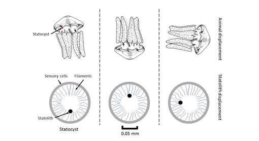
Two of the most important sensory receptors in sea jellies are their eyes and the receptors that allow them to detect their orientation, both of which are generally located around the margin of the bell. The receptors for detecting orientation are called ‘statocysts’. Statocysts are tiny spheres (<1mm diameter) that are lined with nerve receptors and contain a mobile ball (the statolith) that rolls around as the sea jelly changes orientation. As it rolls, it depresses the nerve receptors that line the sphere, which enables the sea jelly to work out whether it is swimming upright, upside-down or horizontally.
Most sea jellies have very simple eyes that can detect the intensity of light but cannot form an image. The box jellies (Class Cubozoa) may be an exception. Their eyes are complex and are very similar in form to human eyes and have a pupil, a lens and a retina. Chironex fleckeri, the most dangerous species of box jelly, has 60 eyes in total! Some scientists think that these eyes enable box jellies to see images. However, images are generally formed and interpreted in the brain and without a brain, or even a complex nervous system, it is unlikely that box jellies can form images like those that we see.
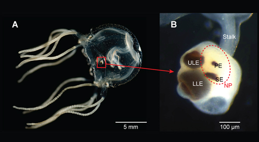 Cubozoan eyes are in structures called rhopalia (A). They have numerous eyes (B) including a lower lens eye (LLE); upper lens eye (ULE), pit eye (PE) and slit eye (SE). The light sensitive neuropil (NP, red broken line) is thought to be involved in diurnal activity. Credit, Bielecki J, Zaharoff AK, Leung NY, Garm A, Oakley TH
